

Lasers for Fluorescence Microscopy
Fluorescence microscopy basics
Over the last decade, fluorescence based life science research has been revolutionized by new imaging methods and the transitioning from bulky gas-laser sources into solid-state lasers with a smaller footprint, longer lifetime, and lower maintenance requirements.
One of the most important tools for microbiology research is high-resolution live-cell imaging through fluorescence microscopy, in which certain molecules, so-called fluorochromes or fluorophores, return low-energy light after excitation with light of a defined wavelength, i.e. light with a higher wavelength than the excitation light. Scientists are taking advantage of this physical effect to investigate ever smaller structures visible in biological processes. Striving to increase the resolution has led to the need not only to use special microscopes but also suitable light sources such as lasers of different wavelengths. In particular, the exact selection of suitable lasers enables the temporal and local resolution to be increased. Lasers are an integral part of modern fluorescence microscopy!
The development of compact, reliable solid-state lasers was an initial enabling technology for commercialization and expansion of high-resolution fluorescence microscopy techniques to new markets and applications, accompanied by parallel improvements in data storage and advanced camera systems, to name a few. While some microscope applications are able to utilize the advancements in LED and super-continuum white-light sources; the high-resolution, high-speed techniques like confocal microscopy still rely on the high-brightness and wavelength precision of lasers.
Modern fluorescence microscopy consists of an enormous variety of different techniques ranging from standard laser scanning confocal microscopy, TIRF and spinning-disc microscopy to light sheet microscopy and various approaches for super-resolution imaging using spatial manipulation of the fluorescence signal.
All these techniques put many different demands on the excitation sources being used, in terms of wavelengths, power levels, power modulation, beam quality and spectral characteristics. A common factor across most techniques is the typically need for many excitation wavelengths in order to address a continuously increasing number of fluorophores and to achieve multi-color imaging.
Most microscope set-ups with multi-color excitation capability typically use multiple individual lasers combined through optical elements and coupled into one or more output beams or optical fibers. Such a laser combiner offers the greatest flexibility in all respects, many wavelengths, many power levels, as well as fast and slow modulation. However, for systems and set-ups where flexibility is not the highest priority, a multi-line laser can offer a more permanently aligned, easier to use and maintenance free alternative.
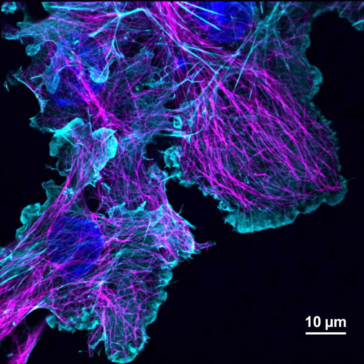
An example of an image taken in a single-molecule localization microscopy
(SMLM) setup including Cobolt Skyra™ from Department of Biotechnology
& Biophysics at Julius-Maximilian-University of Würzburg.
Single and multi-line lasers for fluorescence microscopy
At HÜBNER Photonics we have a very large selection of single and multi-line lasers by Cobolt, perfect for fluorescence microscopy applications. The fast modulation capabilities of our Cobolt 06-01 laser diodes in particular make them very suited to all confocal systems such as cLSM, Spinning Disc and TIRFM. In addition, the excellent on/off modulation, including true off, make them even more attractive for applications such as Photoactivation, Photoconversion, Optogenetics, Laser manipulation, FRET, FRAP, to name just a few. In addition, they are also suitable for super-resolution microscope techniques such as STORM, PALM and STED, since these lasers are available over a wide wavelength range even with high powers.
All Cobolt lasers can be integrated into very flexible and user-friendly laser combiner solutions on the C-FLEX platforms with up to 8 lasers in one combiner. The unique C-WAVE laser offers wide tunability across the almost the complete visible and near IR spectrum, enabling access to more exotic fluorophores. In addition, we can offer the Cobolt Skyra, which is an extremely compact, permanently aligned, multi-line laser that simplifies the integration of multi-color excitation in compact microscopy equipment by eliminating the need for in-field alignment and service, which reduces manufacturing cost and allows for more compact designs. The Cobolt Skyra can also be configured to make a perfect compact solid-state alternative to Ar-ion lasers.
Recent laser-based imaging methods try to combine different techniques such as cLSM and Raman spectroscopy. The Cobolt lasers of the 04/05 and 08-01 Series are well suited for Raman spectroscopy due to the very narrow linewidth and excellent spectral purity. Our C-FLEX laser combiner is the perfect solution to combine both Cobolt 06-01 Series of plug & play diode lasers and the narrow linewidth lasers from the 04/05 and 08-01 Series, thus offering maximum flexibility for many Life science applications.
Understand where our lasers and combiners are used in life science applications in our webinars:
Multi-Line Lasers or Laser Combiners: What Solution Is Best for Fluorescence Imaging?

Watch our webinars and Laser Lounge sessions.
Browse through our dedicated playlist


Cobolt 06-01 Series
Plug & play modulated CW lasers
Wavelength: 375 nm – 1060 nm
Power: 40 mW – 400 mW
Applications: Microscopy, flow cytometry, optogenetics

Cobolt Skyra™
A revolutionary multi-line laser platform
Wavelength: 405 nm – 685 nm
Power: 50 mW, 100 mW
Applications: Microscopy, flow cytometry
C-FLEX
The compact and flexible laser combiner
Wavelength: 375 nm – 1064 nm
Power: 50 mW – 1000 mW
Applications: Microscopy, Raman, holography
Cobolt Rogue™ Series
High Power, CW diode pumped lasers
Wavelength: 640 nm
Power: 1 W
Applications: Super resolution microscopy

VALO Femtosecond Series
Ultrafast femtosecond fiber lasers
Pulse duration: Down to 30 fs
Power: Up to 3 W
Applications: Multiphoton Microscopy, Two-photon Polymerization, Optogenetics
Cobolt 04-01 Series
Single frequency, CW diode pumped lasers
Wavelength: 457 nm – 1064 nm
Power: 25 mW – 400 mW
Applications: Raman, microscopy, LDV, DLS
Cobolt 05-01 Series
High power, single frequency, CW diode pumped lasers
Wavelength: 320 nm – 1064 nm
Power: 10 mW – 3 W
Applications: Holography, Raman, microscopy, flow cytometry, research
Editorial STORM made easy for high-throughput research | Laser Focus World – BioOptics Aug 2021
Publication M. Haahr et al. Permanently aligned multi-line lasers SPIE 2020 Paper 11231
Publication T. Grotjohann et al, rsEGFP2 enables fast RESOLFT nanoscopy of living cells eLife 2013
Publication S. Doose et al Single molecule FRET. Biophysics Letter 2007
Publication M. Dueser et. al Single molecule FRET SPIE Vol 6092 2006
Publication N. Zarrabi et. al. Monitoring the rotary motors of single FF-ATP
Publication U. Krzic et al. Cobolt Mambo 594nm for 3D imaging of live specimen (German version)
Editorial Multiline lasers for fluorescence microscopy PhotonicsViews June 2019
Editorial Highly stable multi-line lasers for next-generation imaging BioOptics 2018
Editorial CW DPSS Lasers make STED Microscopy More Practical BioPhotonics 2012
Editorial Improved contrast with 473nm for confocal microscopy OSA CLEO CThII5 2003
White paper Multi-line lasers simplify biomedical imaging
Poster Novel 515 nm DPSS laser. Focus on Microscopy 2008
Apps note Cobolt Zouk 355 nm for fluorescence microscopy and flow cytometry
Apps note TRAST microscopy for measuring oxygen concentration
Apps note Cobolt Flamenco™ 660 nm for Stimulated Emission Depletion (STED)
References
Literature 04-01
| References | Application | Publication |
| A. Tengholm et al. Fluorescent protein vectors for pancreatic islet cell identification in live cell imaging | TIRF microscopy | Pflügers Arkiv 468:1765-1777 (2016) |
| S. Barg et al. Contact-induced clustering of syntaxin… | TIRF microscopy | Nature Communications 5:3914 (2014) |
| C.Hertlein et.al. TIRF microscopy | TIRF microscopy | ACS 2007 |
| Cobolt Mambo 594nm for 3D imaging of live specimen | 3D Imaging | Photonik International, 2011 |
| Cobolt-Mambo-594nm-for-flow-cytometry | Flow cytometry | Application note |
| Cobolt-Mambo-594nm-for-Raman | Raman spectroscopy | Application note |
| E.Illy et.al Low noise orange lasers for flow cytometry | Flow cytometry | CYTO 2010 |
| Fluorescence microscopy of living cells 515 nm lasers | Fluorescence microscopy | Photonik International Magazine 2009 |
| G.Karlsson et.al. Improved contrast with 473nm | Confocal microscopy | OSA CLEO CThII5 2003 |
| K.Peneva et. al. FCS | Fluorescence microscopy | J. Am. Chemical Society 2007 |
| L.Kaestner et.al. Novel 515 nm DPSS laser | Microscopy | Focus on Microscopy 2008 |
| Multiline DPSS lasers-true Ar+ alternative | Fluorescence microscopy/ Flow cytometry | Europhotonic, July 2004 |
| M. Duser et. al Single molecule FRET | FRET microscopy | SPIE Vol 6092 2006 |
| N. Zarrabi et. al. Monitoring the rotary motors of single FF-ATP | FRET microscopy | |
| Orange Laser Sources for Life Sciences Research | Fluorescence microscopy/ Flow cytometry | BioPhotonics Magazine, Jan 2010 |
| S.Doose et. al. Single molecule FRET. | FRET microscopy | Biophysics Letter 2007 |
| T.Grotjohann et al, rsEGFP2 enables fast RESOLFT nanoscopy of living cells | RESOLFT microscopy | 2013 |