

31 July, 2025
Femtosecond lasers for multiphoton microscopy
Multiphoton microscopy has transformed biological imaging by enabling high-resolution, three-dimensional visualization without the need for dyes or labels. Central to this advancement is the use of ultrashort femtosecond laser pulses. These short pulses minimize light exposure, significantly reducing photodamage during imaging. This is especially critical when studying live samples, as it allows researchers to capture detailed images while preserving the sample’s viability.
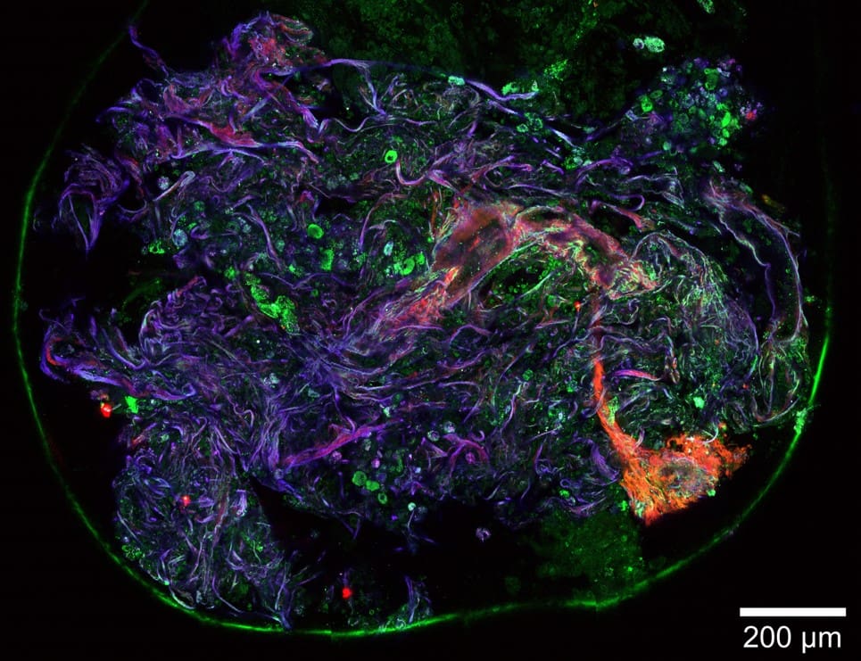
Multi photon microscopy image of lung tissue. Co. of Prof ML Groot Vrije Uni Amsterdam.
Ultrafast femtosecond lasers, with their significantly shorter pulse durations compared to conventional systems, deliver much higher peak power while maintaining low average power. This combination enhances imaging contrast and reduces heat exposure to the sample, which is crucial for preserving biological integrity during imaging. The short pulse duration minimizes photodamage and photobleaching, making these lasers ideal for long-term studies of living cells and tissues.
The high peak power of sub-40 femtosecond pulses is essential for driving nonlinear optical processes such as two-photon excitation (2PE), second-harmonic generation (SHG), three-photon excitation (3PE), and third-harmonic generation (THG), whose signal intensities scale with the square or cube of the incident light intensity. These advanced techniques enable precise, label-free imaging of molecular and structural features with exceptional clarity.
Second-harmonic generation, a coherent nonlinear process, occurs in non-centrosymmetric structures like collagen fibrils, microtubules, and other ordered biological assemblies. This makes SHG particularly valuable for imaging connective tissues, cytoskeletal elements, and fibrillar proteins with high specificity and contrast. In contrast, third-harmonic generation arises at interfaces with refractive index mismatches or in regions of material inhomogeneity, offering detailed visualization of cell membranes, lipid-rich domains, and organelle boundaries.
By integrating these nonlinear imaging modalities, researchers can extract rich, multidimensional information from biological samples—enabling deeper insights into cellular architecture and function.
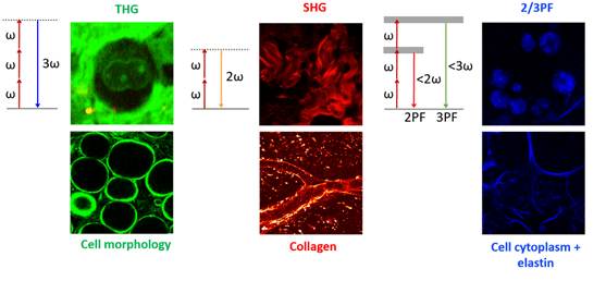
Fig 2: Example of THG,SHG and 2 and 3 Photon Flouresence, simultaneously detected in different channels, outlining the different parts of the biological samples which can be investigated depending on the nonlinear optical technique chosen.
Three-photon efficiency with short pulse lasers
Since the efficiency in multiphoton excitation is strongly dependent on the peak power of the incident light during the pulse, the shorter the pulse, the higher the peak power and the stronger the generated multiphoton signal. Up till now there has been relatively limited ability to generate ultrashort and clean pulses below the 80 fs limit.
The VALO Femtosecond Series offer a novel approach to overcome this limit, resulting in much shorter (sub 50 fs) and clean pulses. Figure 3 illustrates the relationship between the two-photon and three-photon efficiencies and the pulse duration [1, 2]. The efficiency of multiphoton events depends non-linearly on the laser peak power, and the corresponding signal increases with the square and cube of the incident light’s peak power, for two and three photon processes, respectively. For instance, decreasing the pulse duration from 200 fs to 50 fs results in a 4 times higher peak power, the two-photon efficiency is increased by a factor of 4 while the three-photon efficiency increases by a factor of 16.
Another advantage of using femtosecond pulses is increased efficiency of two-photon fluorescence. The high peak power of these pulses enhances the nonlinear interaction, resulting in increased fluorescence efficiency. This allows researchers to obtain high-resolution images with fewer photons, reducing the need for high laser power and minimizing photodamage.
In addition, femtosecond pulses provide improved spatial resolution in multiphoton microscopy. The short pulse duration and high peak power allow for high-resolution imaging in 3D, enabling researchers to study samples at the cellular and subcellular level.
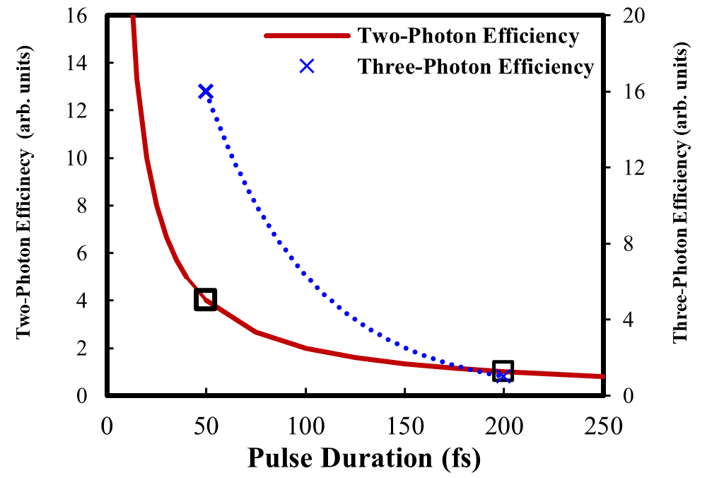
Fig. 3: Two and three photon efficiency vs. pulse duration
In essence the shorter laser pulses mean higher peak intensity, which dramatically boosts imaging signals:
- Two-photon efficiency (2PE) signals increase 5-fold with shorter pulses.
- Three-photon efficiency (3PE) signals can increase up to 25 times.
These gains not only improve image quality but also reduce heat generation and photobleaching, making long-term imaging of live cells safer, more stable, and more effective.
Why sub-50 femtosecond pulses are critical for three-photon imaging
To experimentally validate the benefits of shorter laser pulses, it is crucial to accurately measure pulse duration at the sample. Without proper dispersion compensation in the microscope optics, pulses become stretched, reducing peak power at the focal point. This compromises the efficiency of higher-order harmonic generation and makes experimental verification unreliable.
To generate strong nonlinear signals—such as second-harmonic generation (SHG) and third-harmonic generation (THG)—while maintaining a gentle average power suitable for live samples, sub-50 femtosecond pulses and effective dispersion pre-compensation are essential. These conditions ensure a sufficient signal-to-noise ratio without compromising sample integrity.
Figure 4a) shows the third harmonic signal from a calibration grid using an average power of 4.7 mW on the sample, with the full bandwidth of the VALO Femtosecond Series laser, yielding ~40 fs pulses. In Figure 4b), the spectral bandwidth of the laser was restricted to 10 nm FWHM around 1 064 nm, resulting in ~160 fs pulses. The image in Figure 4a) and 2b) are identically scaled, but Figure 4b) shows no THG signal. Only after rescaling the lower THG signal from the longer ~160 fs pulses, was it possible to obtain an image above the experimental noise floor, as shown in Figure 4c). In this case, a 2.5 times higher average laser power was needed to achieve a signal to noise ratio comparable to that of THG signals obtained from shorter sub 50 fs pulses [3].

Figure 4: Third harmonics of a calibration grid (Ibidi) with 50 micrometer squares. a) 4.7 mW, full spectrum short pulses (<50 fs; VALO Series). b) 6 mW with laser spectrum limited to 10 nm bandwidth (~160 fs). c) Scaled up contrast for the 6 mW laser spectrum limited to 10 nm bandwidth (~160 fs).
Advanced Three-Photon Microscopy with ultrafasr lasers
Compared to convetional femtosecond laser systems, VALO ultra-short pulse lasers generate a much broader optical spectrum up to 1140 nm, reaching deeper into the near-infrared (NIR) range (see Fig 5). his expanded bandwidth enables excitation not only of two-photon processes such as second-harmonic generation (SHG) and two-photon excitation (2PE), but also of more advanced nonlinear phenomena like three-photon excitation (3PE) and third-harmonic generation (THG).
This is possible because the generated three photon signal can efficiently pass through standard microscope objectives as it will not be blocked by the transmission of a standard microscope objective. Therefore, the infrared spectrum needs to cover wavelength ranges above 1080 nm. In contrast, traditional laser wavelengths around 1 µm produce a THG signal in the UV range, which is often too short to be transmitted through standard objectives. Quite simply, the very broad optical bandwidth of these new laser systems allows for simultaneous 2PE, SHG, THG and 3PE generation and detection.
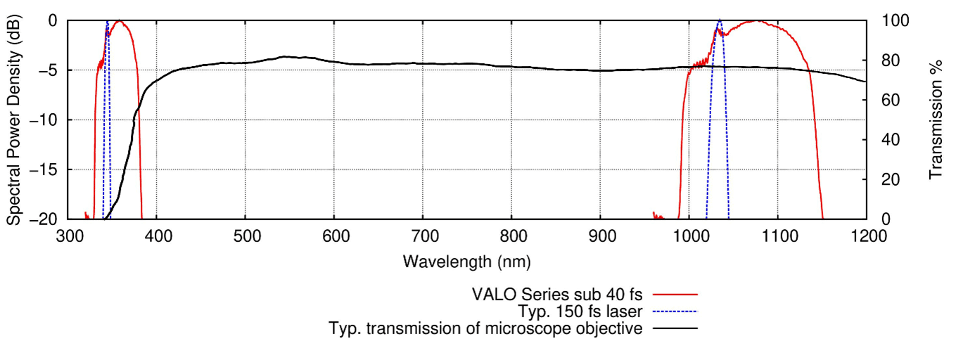
Fig 5: Fundamental and third harmonic generation spectrum of a 30 fs, broadband fiber laser (red) compared with standard 150 fs lasers and typical transmission characteristics of a standard microscopy objective (black solid line). Only a THG spectrum generated from wavelengths of above 1080 nm will be transmitted
Real-Time Cancer Diagnosis with Multiphoton Microscopy
One of the most promising applications of multiphoton microscopy is in cancer research, and specifically cancer diagnostics. Traditional biopsy analysis can take days, but higher harmonic generation (HHG) imaging can deliver results in minutes. A recent study showed that a biopsy could be imaged and analyzed just six minutes after excision, with 87% accuracy [4] .
This technique is especially valuable during procedures like bronchoscopy, where obtaining sufficient tissue for diagnosis is challenging. HHG imaging provides immediate feedback, potentially eliminating the need for repeat procedures.
Start-up Flash Pathology, spun out of Vrije Universiteit Amsterdam, is developing a compact HHG microscope for clinical use. Their goal? To bring real-time, label-free tissue imaging into hospitals, revolutionizing diagnostics. This is possible because they integrated our VALO femtosecond laser into a mobile microscope that can be transported directly into the surgery room.
Watch in this webinar as Dr. van Huizen from Flash Pathology and Dr. Oliver Prochnow outline the real-life applications of multiphoton microscopy and higher harmonic generation imaging.
Conclusions
In summary the use of very short femtosecond pulses in multiphoton microscopy has several advantages, including reduced photodamage, increased efficiency, improved spatial resolution, and reduced background signal. In particular, the ultrashort sub 50 fs pulses provide considerably higher pulse peak powers, which results in optimal signal-to-noise ratio images at much lower average power, which in turn reduces photobleaching, and extends cell viability. These advantages have made multiphoton microscopy a valuable tool in the study of biological samples, allowing researchers to obtain high-resolution images with minimal photodamage.
References
[1] Shuo Tang, Tatiana Krasieva, Zhongping Chen, Gabriel Tempea, Bruce Tromberg (2006), Effect of pulse duration on two-photon excited fluorescence and second harmonic generation in nonlinear optical microscopy, Journal of Biomedical Optics, 11(2).
[2] Mira Sibai, Hussein Mehidine, Fanny Poulon, Ali Ibrahim, M. Juchaux, J. Pallud, A. Kudlinski, Darine Haidar (2018), The Impact of Compressed Femtosecond Laser Pulse Durations on Neuronal Tissue Used for Two-Photon Excitation Through an Endoscope, Scientific Reports, 8:11124.
[3] White paper, Sub 50 femtosecond pulse lasers for gentler multiphoton microscopy, HÜBNER Photonics. In publication Feb 2023.
[4] Prochnow Oliver, van Huizen Laura (2025), Unveiling cell secrets with lasers, Photonics Views.
………………………………………………………………………………………………………………………………………………………………………………………………..
Learn more about the range of ultrafast femtosecond fiber lasers from HÜBNER Photonics. With outstandingly high pulse peak power levels and computer controlled group velocity dispersion pre-compensation, the VALO femtosecond fiber lasers are ideal for multiphoton imaging.

VALO Femtosecond Series
Ultrafast femtosecond fiber lasers
Pulse duration: <40 fs
Power: Up to 3 W
Applications: Multiphoton Microscopy, Two-photon Polymerization, Optogenetics
More resources
Explore our Publications for practical insights on how our customers are leveraging the power of our lasers in their projects.

Customer publications
Application: Holography
Product line: Cobolt
Wavelength: 405 nm
Cloud Measurement with SmHOLIMO Holographic Imager
SmHOLIMO (Small Holographic Imager for Microscopic Objects), a cutting-edge holographic imager designed for high-resolution in situ measurements of cloud droplets.
Read summary of article "Cloud Measurement with SmHOLIMO..."

Our publications
Application: LIBS
Product line: Cobolt
Wavelength: 1064 nm
High-resolution High-Speed LIBS Imaging
This work demonstrates an approach to reduce the acquisition time for high resolution µ-LIBS imaging by using a laser operating in the kHz frequency range.
Read summary of article "High-resolution High-Speed LIBS..."

Customer publications
Application: LIBS
Product line: Cobolt
Wavelength: 1064 nm
Denoising Approaches in LIBS Imaging
The researchers evaluate and compare 5 denoising methods with the objective of enhancing SNR (signal-to-noise ratio) in fast μLIBS imaging.