

New Editorial in Wiley’s Photonics Views Magazine Jan 2020 edition:
Digital holographic microscopy: Requirements of lasers for a novel label-free cytometry tool
by Björn Kemper & Elizabeth Illy
[Extract] Digital holographic microscopy (DHM)[2,3] is an interferometry-based variant of QPI (quantitative phase imaging) in which typically a laser is used as a coherent light source. DHM provides QPI by detecting specimen-induced optical path length changes against the surrounding environment. The specific requirements of the laser depend on the application and the samples under investigation, however the absolutely most important requirement is long coherence length and small footprint. DHM can be modularly integrated into common optical microscopes [3,4]. This allows effortless integration and usage of the technology as a label-free imaging tool in research laboratories. Read the whole article for free at Photonics Views
In DHM, reconstruction of digitally captured holograms is performed numerically with a computer. This feature allows imaging of different image planes of a specimen (multifocus imaging) without mechanical focus realignment5 and even subsequent autofocusing after capturing of digital holograms is possible. In this way unpredictable focus drifts, e.g., due to environmental condition changes during long-term observations of living cell cultures, or caused by specimen induced movements inside 3D environments such as collagen or gel matrices, can be efficiently corrected. DHM-based QPI moreover allows sophisticated and robust automated object tracking for determination of cell migration and motility and enables in combination with state of the art image processing like segmentation to extract biophysical specimen parameters such as volume, thickness, and dry mass1. In addition, also optical parameters such as the cellular refractive index, which is related to water content, intracellular solute concentrations, and tissue density3,6 are accessible from DHM quantitative phase images. These features qualify DHM as a versatile label-free in-vitro imaging tool to analyze tumor, blood, and stem cells in mixtures or composites and allow to study their responses to chemicals or drugs.
Lasers for DHM
The most important performance parameter requirement on a laser for DHM is the coherence length. By coherent this mean that all the light waves travel in synchronization i.e. they have the same period and phase, and this characteristic is found in truly single longitudinal mode (SLM) or single frequency (SF) lasers. The coherence length of a light source is directly correlated to the spectral linewidth of the emitted light (temporal coherence), as well as the homogeneity of the phase front over the beam cross section (spatial coherence). The distance the light needs to be coherent over in order to make an interference pattern is determined by the depth of field; the larger the depth of field the longer the coherence length that is needed. Regularly a coherence length of >1m is more than sufficient. However, typically a larger coherence length allows for more complex and flexible holographic setups.
Besides the coherence length, there are a few other parameters which are important to be considered when selecting a laser for DHM. The wavelength is not so critical so other visible colours, like 561 nm, or near infrared (NIR), e.g., typical Raman wavelengths, could also be used, and may be favorable for specific applications. The main considerations for the wavelength are camera sensitivity (eg CMOS and CCD) and sample properties. For example, short wavelengths in the visible spectrum (VIS) allow QPI with high interferometric sensitivity and latera resolution and are well suited for investigations on thin biological objects such as cells in a Petri dish, while light in the NIR, e.g. 785 nm, is advantageous for imaging thicker samples such as tissue slices. Direct light modulation at up to 10s Hz frequencies, as needed, for example, to minimize the light exposure to living biological specimens during time-lapse investigations, can be achieved with external devices such as AOMs however these add complexity and space, something to be considered when aiming to commercialize the system. A directly modulated compact SLM laser is therefore highly attractive. Since fiber delivery simplifies the design, the beam pointing stability is important to avoid power fluctuations at the sample, as is the beam profile in order to maximise coupling efficiency (Figure 2a).
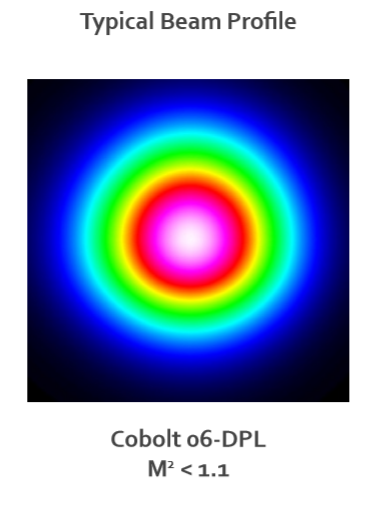
Fig 2a: Typical beam profile of the SLM 06-DPL used for DHM.
Meet the authors
Björn Kemper, Ph.D., is a senior researcher the Biomedical Technology Center of the Medical Faculty at the University of Münster, Germany. Email: bkemper@uni-muenster.de.
Elizabeth Illy, Ph.D., is the Head of Marketing at HÜBNER Photonics. Email: Elizabeth.illy@coboltlasers.com
References
1. Y.K. Park et al. (2018). Quantitative phase imaging in biomedicine. Nat Photon, Vol. 12, pp. 578-589.
2. B. Kemper and G. von Bally (2008). Digital holographic microscopy for live cell applications and technical inspection. Appl Opt, Vol. 47, pp. A52-A61.
3. B. Kemper et al. (2019). Label-free quantitative in-vitro live cell imaging with digital holographic microscopy. In Bioanalytical Reviews, J. Wegener, Ed. Springer Nature Publishing: Basel, Switzerland.
4. P. Lenz et al. (2016). Multimodal quantitative phase imaging with digital holographic microscopy accurately assesses intestinal inflammation and epithelial wound healing. J Visualized Exp, (115) e54460.
5. P. Langehanenberg et al. (2011). Autofocussing in digital holographic microscopy. 3D Res, Vol. 2, Issue 4.
6. D. Bettenworth et al. (2018). Quantitative phase microscopy for evaluation of intestinal inflammation and wound healing utilizing label-free biophysical markers. Histol Histopathol, Vol. 33, Issue 5, pp. 417-432.
7. L. Kastl et al. (2017). Quantitative phase imaging for cell culture quality control. Cytom A, Vol. 91, Issue 5, pp. 470-481.