
Interferometric Image Scanning Microscopy for Live Cells
Researchers at Stanford University have unveiled a powerful new microscopy method that promises to transform the study of living cells.
A team led by Michelle Küppers and W. E. Moerner (recipient of the 2014 Nobel prize in Chemistry) has developed interferometric image scanning microscopy (iISM), a technique capable of imaging structures inside live cells with ~120 nanometer resolution, while significantly reducing light-induced damage.
Conventional fluorescence imaging can suffer from phototoxicity, limited labeling efficiency, or perturbation of biological function. Other label-free approaches typically struggle with low contrast and limited resolution in complex cellular environments.
The new approach lies in combining interferometric scattering microscopy, which detects nanoscale structures based on their light scattering without labels, and image scanning microscopy (ISM), a technique that enhances resolution and signal quality using detector arrays and computational reconstruction.
By merging these approaches, iISM achieves high-resolution, high-contrast imaging while remaining non-invasive.
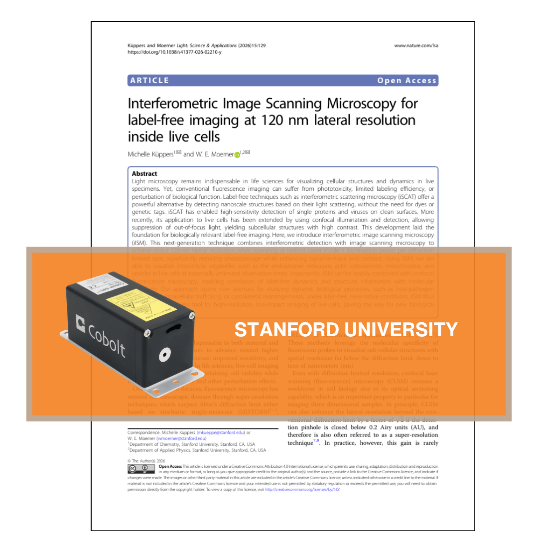
To realise this, the team built a custom ISM microscope capable of both interferometric scattering and fluorescence detection. Illumination was provided by the Cobolt 06-01 MLD diode laser at 445 nm with 150 mW output power, serving as a stable and coherent light source.
Using ilSM, the researchers were able to visualise intracellular organelles and vesicles in live cells at essentially unlimited observation times. Importantly, the technique can be seamlessly combined with confocal fluorescence microscopy, enabling correlation of label-free dynamics and structural information with molecular specificity.
Together, these capabilities position iISM as a powerful new tool for high-resolution, low-impact imaging of live cells, opening the door to deeper insights into dynamic biological processes.
More resources
Looking for more in-depth information? Visit our Knowledge Bank page for detailed articles and insights on our products and technologies.