

World’s fastest 3D Imaging Flow Cytometry System
Researchers at the University of Tokyo have developed a novel method for high-speed 3D imaging flow cytometry using an innovative technique called Optofluidic Spatial Transformation, as detailed in Biomedical Optics Express.
Conventional imaging flow cytometers typically produce only 2D images, limiting the ability to study complex cellular structures. To overcome this challenge, the team developed a fluorescence technique called FLITS (Fluorescence Imaging using Light-sheet Illumination and Tilted Spatial Transformation). By combining laser-based light-sheet excitation with a slightly tilted microfluidic channel, FLITS captures multiple fluorescence slices that are computationally reconstructed into detailed 3D images.
For the FLITS setup the team used the Cobolt 06-MLD 488 nm laser, which provided the excitation for the strobe light-sheet imaging system.
This approach eliminates the need for mechanical scanning, opening new possibilities for high-throughput needed for flow cytometry applications.
 Go back to all publications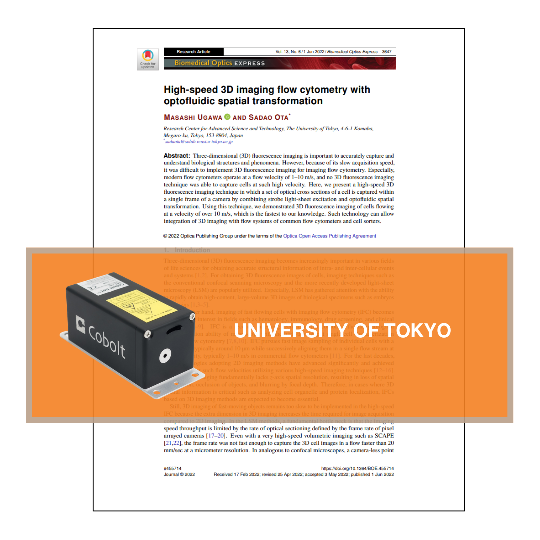
More resources
Looking for more in-depth information? Visit our Knowledge Bank page for detailed articles and insights on our products and technologies.